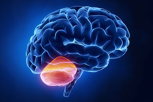
As a follow-up to my last article on functional neuroanatomy and neurological examination techniques [July issue], I want to discuss the cerebellum. When patients present with tremors or ataxias, we commonly label these as Internal Wind and recognize the role of the cerebellum in such cases; however, we were never taught how to clinically assess Internal Wind. Let's explore examinations specific to the cerebellum as a functional means to tracking Wind.
Appreciating the Value of Cerebellar Function Testing
Common knowledge relegates the cerebellum to the seemingly basic task of coordinating movement, something we generally take for granted. A deeper dive into neuroscience reveals the secret life of the cerebellum, quietly orchestrating behind the scenes not only movement, but also learning, memory, emotions, autonomic functions and even hormones.
Considering all these roles, testing cerebellar function becomes important for patients with a wide variety of signs and symptoms including balance deficits, falls, gait abnormalities, muscular imbalances, concussions, traumatic brain injuries, stroke, neurodegenerative disorders, psychiatric disorders and learning disabilities.
 There is no single conclusive bedside neurological exam for the cerebellum. Cerebellar testing must be comprehensive due to the complexity of this region of the brain. For example, I have had patients who performed very well on motor coordination tests for the arms, and very poorly on motor coordination tests for the legs. This is possible because there are very distinct regions of the cerebellum devoted to upper-limb coordination and lower-limb coordination. Cerebellar function is not an all-or-nothing phenomenon. Small areas can be affected, while others remain intact.
Understanding the Anatomy and Physiology
The cerebellum is phylogenically divided into three lobes: the flocculonodular lobe, anterior lobe and posterior lobe. The flocculonodular lobe is involved in vestibular functions, mediating eye movements and spinal stability. The anterior lobe is involved in limb coordination and posture. The posterior lobe is from an evolutionary perspective the newest lobe, and parallels the development of the neocortex. It is involved in aspects of motor planning and fine motor movements, such as those required to play an instrument.
At the most basic level, we have the right cerebellum controlling the coordination of movement on the right side of the body, and the left cerebellum doing the same for the left side of the body. The cerebellum promotes extensor tone in the body. There is a significant amount of real estate in the brain devoted to allowing us humans the unique ability to walk, run, play and stand in line for coffee on two feet; and healthy cerebellar function is vital for the unconscious maintenance of proper muscle tone that allows us to do so.
One of the fascinating observations that can be made in the clinic is a difference in the performance of a test based on whether a patient is seated versus standing. For example, in testing the extensor tone of the thumbs, a patient with cerebellar hypofunction may have good engagement of extensor muscles in the thumbs while seated; however, as soon as they stand up and you repeat the same test, one or both thumbs may test weak.
Why would that happen? As soon as the patient stands up, gravity is having a greater influence on the system, and the cerebellum is now working harder to activate proper extensor tone to maintain an upright position. If the cerebellum is weak, its ability to allocate resources to maintaining an upright posture and simultaneously exert healthy extensor tone of the thumbs is compromised.
I have also witnessed a significant difference in performance in other neurological examinations, such as a head thrust test for the vestibular-ocular reflex, depending upon whether the patient was seated or standing. These examples highlight the importance of developing a functional approach to neurophysiology and neurological examination techniques.
Feed-forward, Feedback, and Efferent Copy
The cerebellum operates as an error correction mechanism in the central nervous system. It utilizes feed-forward, feedback and efferent copy mechanisms in order to accomplish this task.
Feed-forward is our visualization and planning pathway. When we decide we want to make a movement, that activates several areas of the brain including the dentate nuclei in the cerebellum, the basal ganglia and motor planning areas in the frontal lobes; and takes into account the environment and situation before executing a movement. If you want to bring a spoon to your mouth, you would need to make different muscle adjustments based on whether that spoon weighed 10 grams versus 10 pounds. These calculations take place in the brain before movement is ever initiated.
If you have ever watched a 1-year-old try to eat yogurt with a spoon, you are watching the cerebellum's error correction mechanism in action. Every time that spoon misses the mark, spilling yogurt all over the child's face and floor, an error signal is generated that goes up the spinocerebellar tract in the spinal cord into the brainstem and then into the cerebellum, providing feedback and informing the cerebellum that the arm overshot the target.
The efferent copy mechanism is like a blueprint for what that arm-to-mouth movement should have looked like, allowing the cerebellum to compare the "feedback error report" with the "efferent copy" to see where the error occurred. With each error message, the cerebellum learns how to more accurately move the arm, and soon, the toddler is eating like a champion. This is how our cerebellum is involved in learning aspects of movement.
Editor's Note: Part 2 of this article appears in the December issue.
Amy Ayla Wolf (formerly Moll), LAc, DOM, DAOM,specializes in neurological disorders, chronic pain, and concussion recovery at her private practice. She's also a faculty member of the Carrick Institute of Clinical Neuroscience and Rehabilitation. For more information visit
.


