Neurological disorders in all age populations are steadily on the rise. As more and more people are living longer, surviving cancer, strokes, and living in an increasingly more toxic environment, we as practitioners need to be prepared for a new era of diseases of the nervous system.
Frontal Lobe Functions
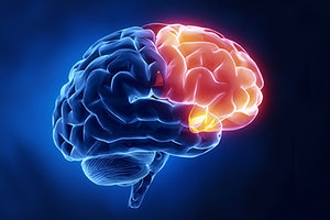
The frontal lobes are responsible for executive functioning, the ability to plan, strategize, problem solve, analyze, and focus attention. They play a major role in emotions, behaviors, and impulse control. These functions are all related to the prefrontal cortex. The frontal lobes have many other distinct regions including the motor cortex, responsible for volitional movements, the frontal eye fields, responsible for initiating rapid eye movements from one target to another (called saccades). Broca's area is found in the frontal lobe and is responsible for the motor aspect of speech. We also have pre-motor areas that aid in the planning of movements. This is why visualization of movements is a powerful tool to prevent atrophy of a damaged limb, or enhance sports performance. Activation of the pre-motor cortex has powerful connections to other parts of the brain, and can affect blood flow to the body.
Testing Eye Movements
 Many patients seeking acupuncture and Chinese medicine have disorders or degenerative diseases of the frontal lobes: depression, emotional imbalances, ADD, ADHD, fronto-temporal dementias, strokes, concussions, and Autism-spectrum disorder to name a few. Other patients may report more subtle deficits in frontal lobe function such as poor concentration, brain fog, cognitive fatigue, and difficulty with planning and organization.
When it comes to working with patients on cognitive symptoms, emotional symptoms, and CNS disorders, it is highly valuable to collect not only subjective data, but also objective findings. This is what neurological exams and cognitive testing can provide. All of these regions of the brain described above have starkly different functions, which is why there is no one test to assess frontal lobe function. It is also important to appreciate that one area of the frontal lobe may be working adequately, while another may not. An excellent example of this is in the testing of eye movements.
From an evolutionary survival perspective, the human with the fastest capability to scan the environment for dangers, is the human that lives. The frontal eye fields allow for the quick initiation of saccade eye movements (rapid eye movements from one target to the next). There are many other areas of the brain that are involved with saccades, however, if we focus on saccade initiation, we get a window into the functioning of the frontal lobe.
The right frontal eye fields allow for a saccade to the left, whereas the left frontal eye field initiate saccades to the right. This contralateral testing is similar for the motor cortex, where the right motor cortex controls volitional movement on the left side of the body. In the human brain, it is possible for differences to be observed in brain output and processing speeds between the right and left frontal lobes. The case of concussion provides a good example of this, where it is often witnessed that neurological examination findings of the right frontal lobe differ from findings on the left.
To perform saccade testing, stand in front of the patient a little more than arms length away. Hold your thumbs up, one centered in front of the patient, and at the level of there eyes. Hold the other thumb approximately shoulder-width out to the patients right. Have them look at the center thumb, and instruct them to look at the lateral thumb as soon as you wiggle it.
Do this 3-5 times, making sure they are not anticipating the movement and jumping to the target ahead of time. Pay attention to how quickly they initiate the eye movement after you wiggle your thumb. Repeat the same test to the left, looking for differences between right and left. Often patients are surprised to discover that they themselves can feel a difference. If a patient has a slower lag-time to initiate an eye movement to the right, this is a sign of deficits in the left frontal eye fields.
The Anti-Saccade Test
In contrast to the saccade, we have another eye movement called an anti-saccade. Anti-saccades provide an assessment window into the prefrontal cortex, the area related to executive functioning. In this test, stand in front of the patient, and hold up two index fingers shoulder-width apart, and in the same plane as your nose (not straight out in front of you). Instruct the patient to look at your nose, and when you wiggle a finger, instruct them to look at the opposite finger that is not moving, then return their gaze to your the nose. Wiggle your fingers in an unpredictable fashion, changing the rhythm as well as pattern. Humans are wired to want to look at visual movement, in order to determine if a sudden movement imposes any threat. In order to accurately perform an anti-saccade test, the patient has to have excellent impulse control, and that requires a healthy prefrontal cortex. Out of 10 times, how many times does a patient accidentally look at the wiggling finger instead of the still finger? If it is more than 3 out of 10, it is likely they are having some difficulty with prefrontal cortical function.
Documenting Improvement Tests such as these are incredibly valuable for serving as biomarkers for patient improvement. Anytime we insert needles into the body we are having an effect on the CNS, however, unless we are assessing the CNS, we have no idea if it is a therapeutic effect or not.
One of the UFC fighters I worked with came to me 2 days following a fight. His saccades had severe latencies both to the left and the right. Following the acupuncture treatment, I rechecked his saccades, and they were remarkably faster. When I see remarkable changes in a neurological exam following a treatment, I know I have been able to do something beneficial for that patient's brain. Another patient came to me for the treatment of a concussion. His saccades were excellent, however he scored very poorly on the anti-saccade test.
This matched up with most of his main complaints, which were all functions related to the pre-frontal cortex. As I treated him, we continued to retest the anti-saccades, and after several treatments, he was able to perform the test quickly with no errors. These changes in nervous system function also coincided with his ability to perform cognitive tasks better, such as lesson planning, as his pre-frontal cortex started to heal.
As we as a profession, start to embrace the first professional doctorate and firmly establish ourselves within the medical profession, we need to be able to objectively document patient improvement, especially in integrative settings where patient charts notes are being shared among multiple providers of different disciplines. We can no longer rely solely on subjective patient information and anecdotal evidence. Neurological and orthopedic exams are an essential step in the evolution of our profession.
Amy Ayla Wolf (formerly Moll), LAc, DOM, DAOM,specializes in neurological disorders, chronic pain, and concussion recovery at her private practice. She's also a faculty member of the Carrick Institute of Clinical Neuroscience and Rehabilitation. For more information visit
.


