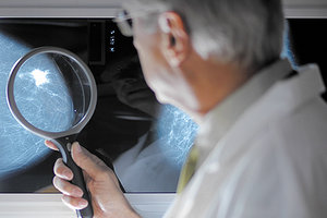
The gold standard test for breast cancer screening in the medical clinics of Western medicine has been mammography for many years. The use of this diagnostic test, however, has always been encumbered with controversy.
Two landmark events have occurred within the past five years that have impacted the controversy over mammography:
1. In 2009, the U.S. Preventive Services Task Force, as a result of an extensive study of the risks versus benefits of mammography, announced its new more stringent recommendations: to begin mammography at age 50, with an exam every two years, instead of every year, continuing up until age 74, then no further mammography after this age.
2. In mid-2013, the Cochrane Commission reported in the New England Journal of Medicine the results of their own highly impartial meta-analysis of the pros and cons of mammography. The Cochrane Commission is perhaps the most highly rated and most highly-respected scientific medical panel in this country, as well as throughout the world. Their study concluded that, due to recent improvements in breast cancer treatment, and the risks of false positives from mammographic breast cancer screening often leading to unnecessary treatment, they stated that "it therefore no longer seems reasonable to recommend breast cancer screening at any age."
 This strong statement was a shock to many physicians, as well as large numbers of patients, especially since the Cochrane Commission is so highly respected. Many are confused and are wondering what to do. This includes many physician friends of mine as well as patients I am acquainted with. We don't know what the U.S. Preventive Services Task Force will do now. For the time being, each woman (in consultation with her treating physician) will have to decide, based on her own unique medical situation, what is the best course of action in terms of whether to go ahead with further mammography or not.
Thermography
In this setting, thermography becomes an especially attractive alternative. This highly refined technology is based on infrared temperature sensing of the breast. Breast tumors radiate heat: they are faster-growing and have more vascularity than the surrounding normal tissue. Medical interest in thermography dates back to 1957, when it was discovered that the skin temperature over a cancer in the breast was higher than that of normal tissue. Thus began a period of extensive clinical use of breast thermal imaging. Studies conducted throughout the 1970s demonstrated the clinical benefits of breast thermography. As early as 1976, at the Third International Symposium on Detection and Prevention of Cancer in New Hampshire, thermography was established by consensus as the highest risk marker for the possibility of the presence of an undetected breast cancer.
Thermography utilizes infrared imaging detection that can measure minute differences in the temperature over all areas of the breast. Images are taken by a special camera without squeezing the breast tissue, as is the case with mammography. After the initial pictures the woman is often instructed to place her hands in a basin of cold water for 60 seconds; then more pictures are obtained. This technique enables expert interpreters of these images to detect breast changes that occur as a result of dysplasia, before actual cancer cells have developed, let alone actual developed cancerous tissues.
Thermography can therefore be utilized to follow breast health on a year-by-year basis. If a woman has areas of increased warmth and a breast biopsy indicates pre-cancerous changes such as dysplasia, the woman can initiate life-style and dietary changes to improve her overall health before any actual cancer has developed.
As thermography is a surface temperature screening technique, many experts have recommended that it should be used in conjunction with mammography, so as to not miss deeper cancers. However with the Cochrane Commission recommending that mammography be discontinued this combined approach remains in question at this time. I personally believe that other imaging studies will replace x-ray mammography, at least in most cases.
Magnetic Resonance Imaging
MRI breast scans are currently available but these are expensive and time-consuming at the current state of this technology. Further advances in breast magnetic resonance imaging will undoubtedly be made so that this approach can become more widely accepted and popular.
Functional MRI scans have been developed that can measure precise differences in blood flow in various parts of human tissues. These fMRI scans are now widely used in brain flow studies, and they can even detect when someone, such as a crime suspect, is lying! When someone is asked a question and they answer by not telling the truth the frontal cortex lights up with more blood flow in areas that do not receive increased flow when the person is telling the truth (you have to consciously think more when you are framing a lie than you do when you are simply telling the truth). These MRI techniques can certainly be applied to detecting the increased blood flow in breast cancers, but so far they are very expensive.
Ultrasound technology has advanced rapidly in the past few years. Now three dimensional color images of the developing baby in utero are widely available. Ultrasound is also widely utilized in diagnosing gallstones and liver and bile duct disease, including cancer of the bile ducts. Further advances in this technology can be applied to breast imaging.
Elastography
Many diseases cause changes in the mechanical properties of tissues. These changes cannot be directly measured by popular imaging devices such as computed tomography (CT), traditional ultrasound (US) and magnetic resonance imaging (MRI).
Elastography is a strain-gauge imaging technique that has been well established in research literature as a promising technology to identify the increased tissue stiffness of an abnormal growth. This technique is an outgrowth of ultrasound imaging.
To acquire an elastography image, the ultrasound technician takes a regular ultrasound image and then pushes on the tissue with the ultrasound elastography transducer to take a compression image. Normal tissue, fibrocystic disease, and benign tumors are typically elastic or soft and will compress easily whereas malignant tumors do not compress at all. This approach to detecting breast tumors needs further refinement but these developments are underway.
Conclusion
Every woman must decide for herself how she wishes to proceed with her own preventative medicine screening, in terms of all aspects of maintaining good health. This includes regular pelvic exams, pap smears and HPV testing when appropriate, routine annual physical exams, blood tests for cholesterol, glucose, C-reactive protein, liver and kidney function, thyroid screening, Vitamin D levels, complete blood counts, urinalysis, and then after age 50, colonoscopy and yearly testing for occult blood in the stool.
All women must also decide what they wish to do about screening for breast cancer. This should involve a thoughtful discussion with their personal physician. The landscape of mammographic screening is changing, as I have indicated above. Women who seek for alternatives will find they have many options such as thermography and the others I have briefly discussed in this article. Good luck to everyone as you enter the portals to your future good health!
Click here for previous articles by Bruce H. Robinson, MD, FACS, MSOM (Hon).


